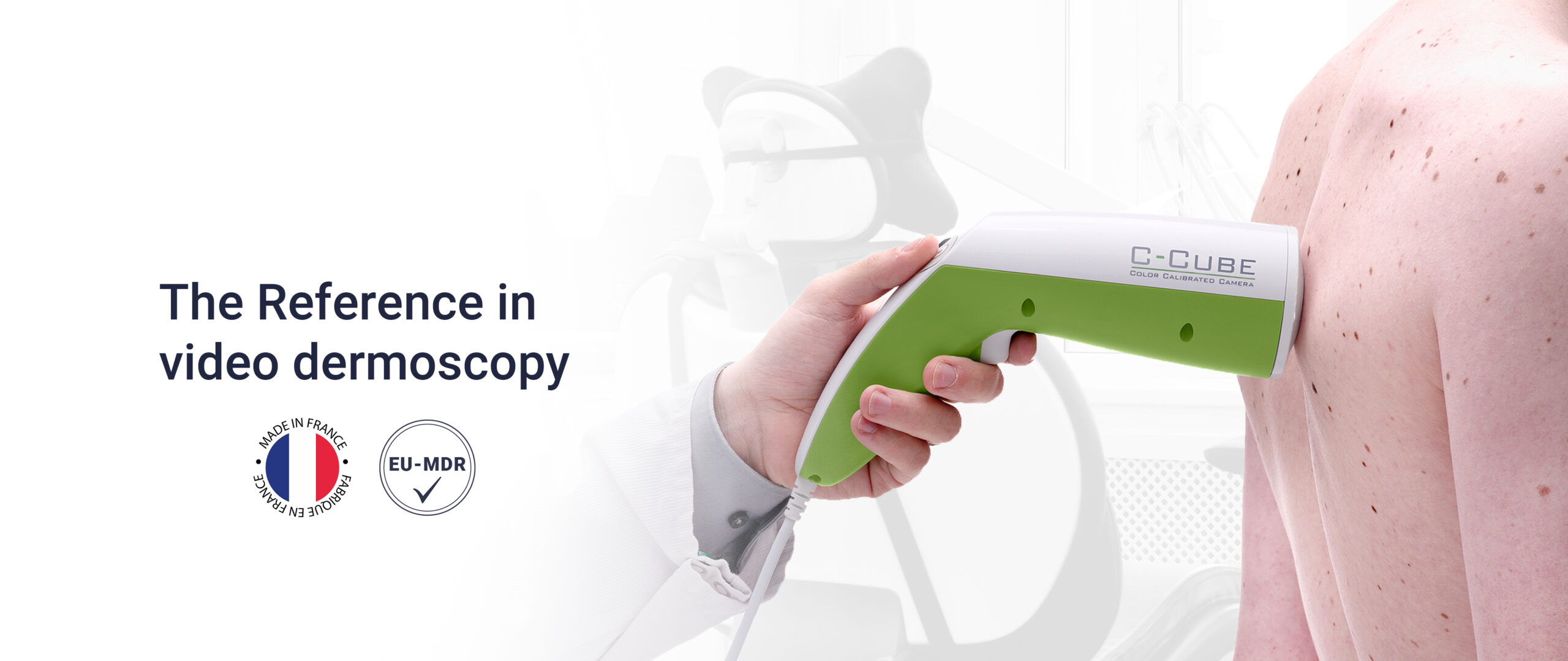
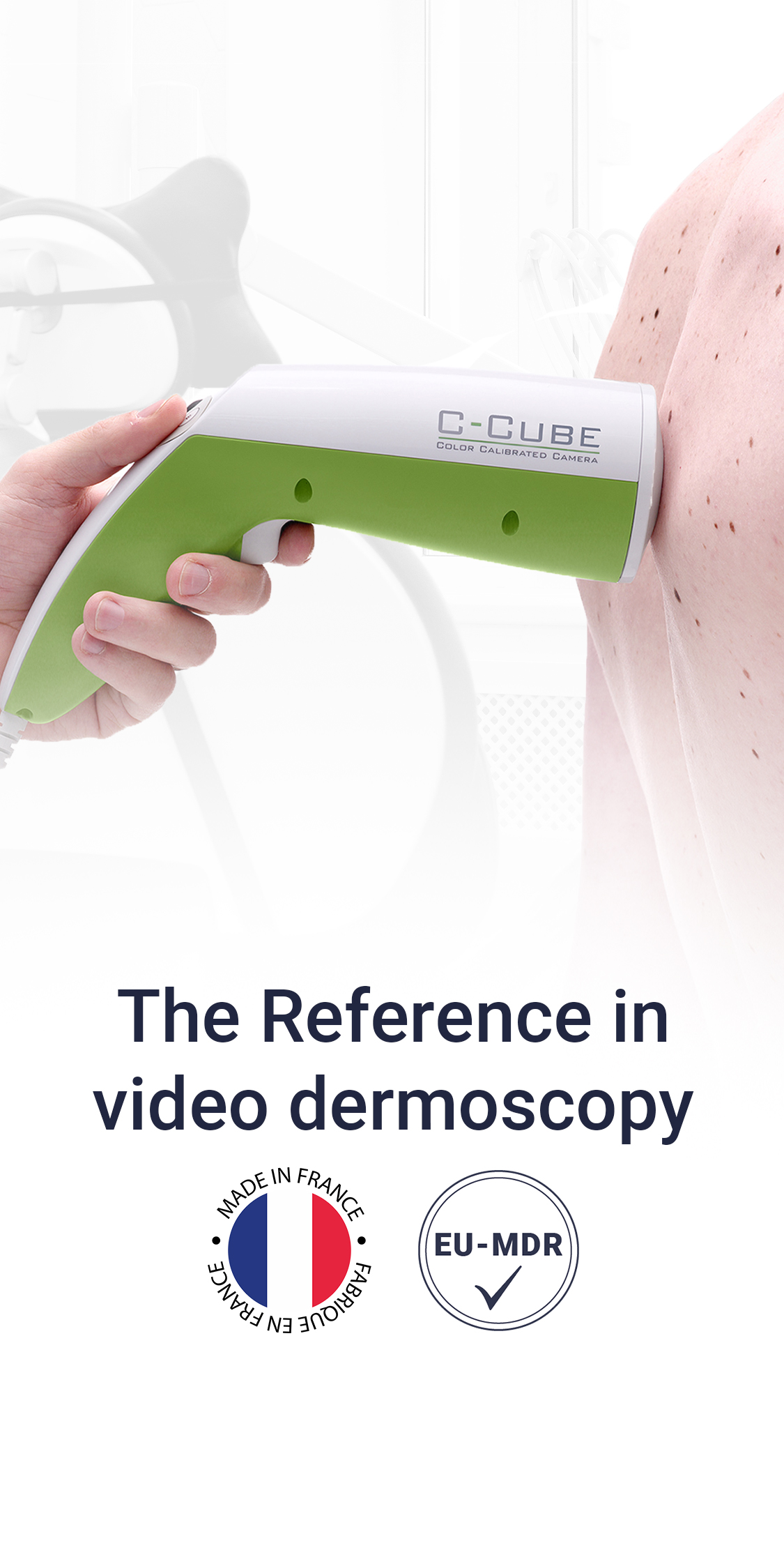
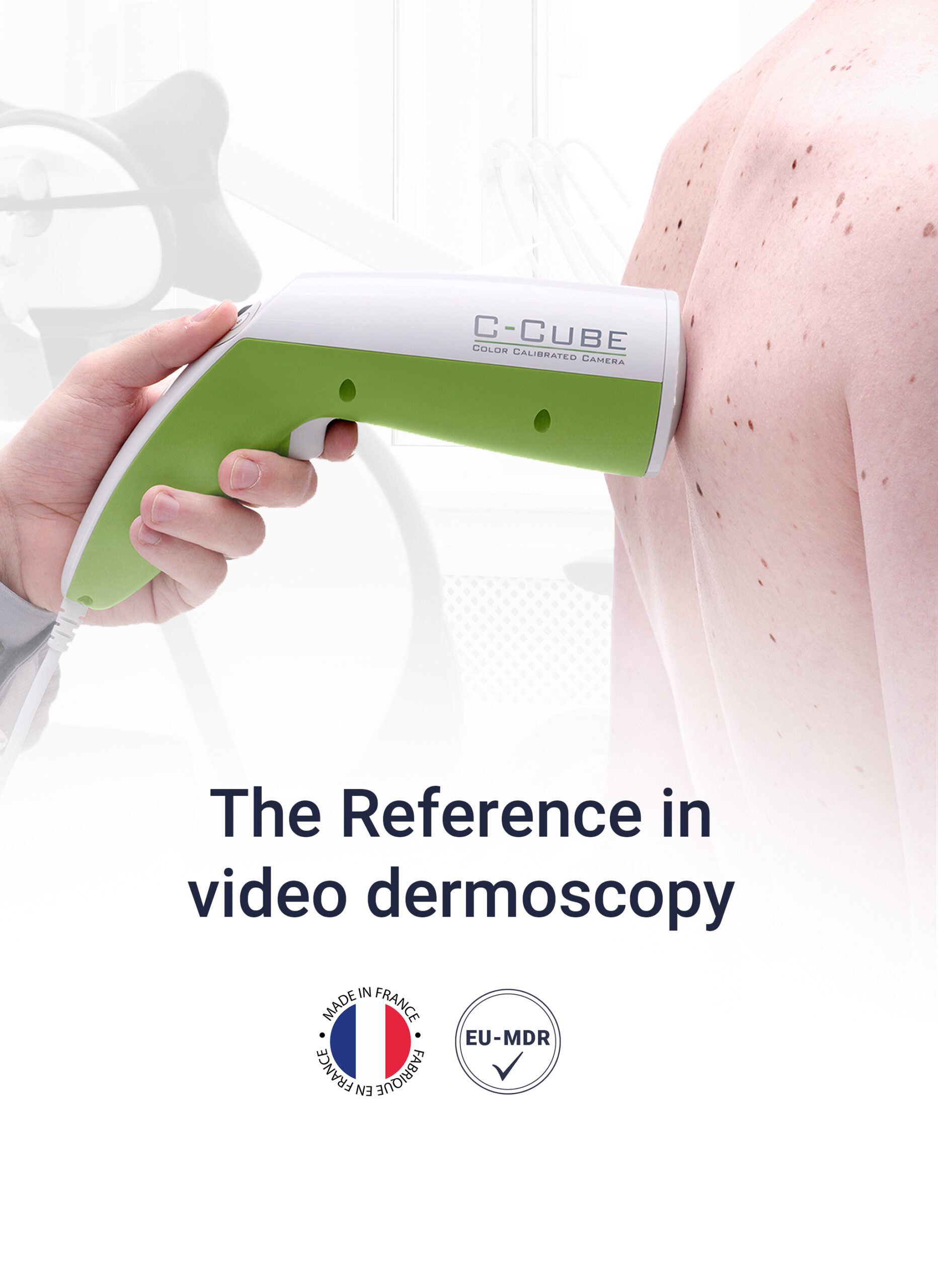
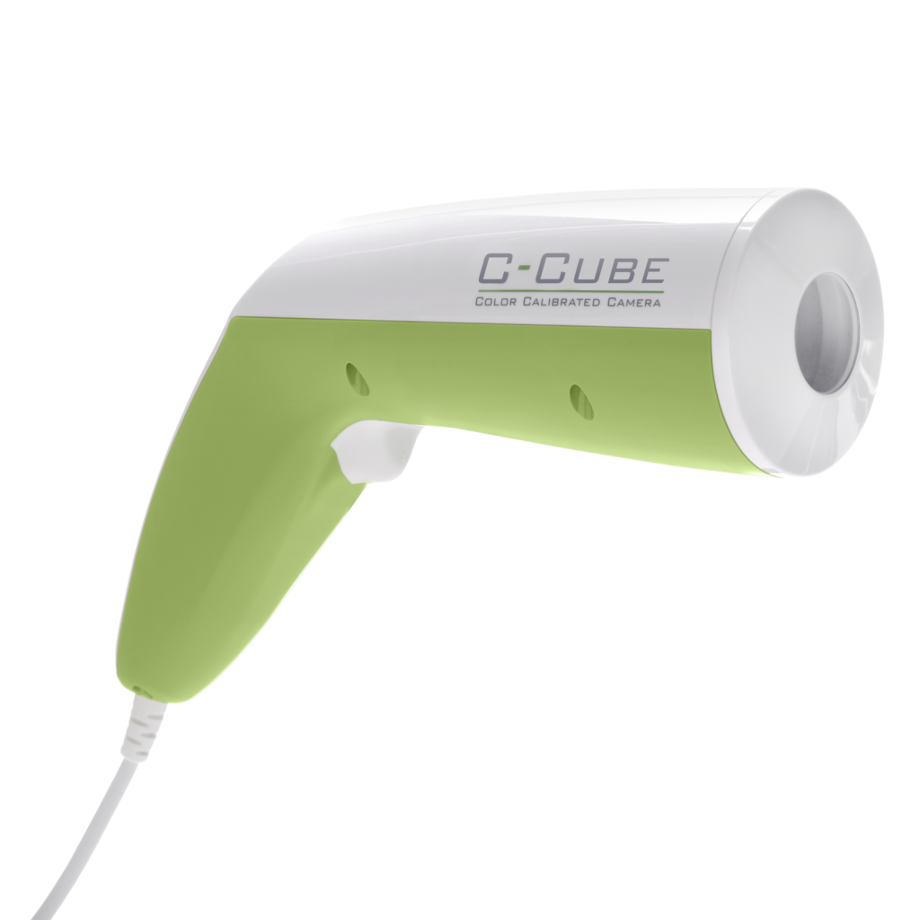
VIDEO DERMOSCOPE C-CUBE 3

Observe. Compare. Monitor​

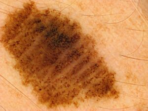
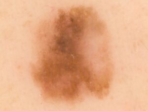
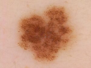
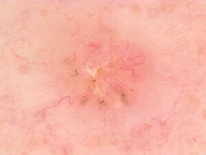
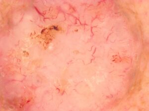
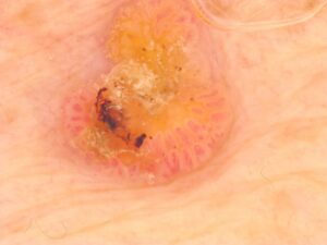
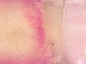
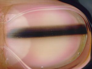
The C-Cube combines Ultra HD video, optical and digital zoom, and TrueColors® technology to enable each lesion to be examined with a level of detail appropriate to the clinical situation.

Thanks to standardised and reproducible imaging, it makes it easier to compare images across consultations and supports dermatologists in monitoring their patients.

A state-of-the-art videodermoscope, designed for monitoring lesions​

The C-Cube has been designed to assist dermatologists in the observation, documentation and longitudinal monitoring of skin lesions.

Its ultra-high-definition dermoscopic imaging allows skin structures to be visualised in great detail. Combined with the patented TrueColors® technology, it ensures accurate and standardised colour reproduction, which is essential for visually assessing how lesions evolve over the course of consultations.

Live on-screen viewing, optical zoom and digital magnification facilitate the examination of lesions and promote communication with the patient.

By building up a history of comparable images, the C-Cube helps to document clinical observations and support dermatological monitoring over time.

Standardised imaging to help make consultations more organised​

The C-Cube fits seamlessly into the day-to-day running of a dermatology practice by making it easier to document lesions and monitor them over the course of consultations.

Thanks to a standardised imaging protocol, photographs can be taken by trained staff, under the doctor’s supervision, allowing the dermatologist to devote more time to the clinical examination, image interpretation and medical decision-making.

This approach facilitates the division of tasks within the practice and enables the photographic monitoring of lesions to be integrated into a structured consultation process.

Reinventing Dermoscopy with a three-dimensional approach

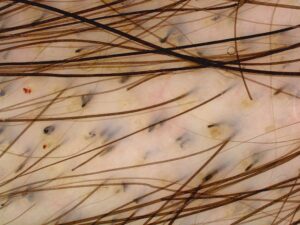
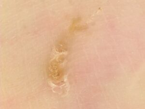
The C-Cube 3 video dermoscope distinguishes itself as a pioneer in digital dermoscopy by incorporating an exclusive feature: the ability to create 3D images.

This technological breakthrough enables visualization of the skin’s relief, offering a new perspective during your medical or aesthetic dermatology examinations.

This capability can also be used to retrieve touch information for remote examinations (tele-expertise).

3D visualisation of a lesion

3D visualisation of a wrinkle

2D and 3D imaging technology documented in scientific publications​

Used for many years in international clinical trials, the C-Cube meets the requirements for reproducibility, standardisation and accuracy expected in the most demanding research protocols. It is the only videodermoscope to have been validated by scientific publications for both 2D and 3D applications.

This scientific expertise contributes to the development of imaging solutions tailored to the observation and monitoring needs of healthcare professionals.

Ergonomy and French Touch

Weighing just 410 g, the C-Cube is the lightest high-end video-dermoscope available, making it easy and comfortable to use.

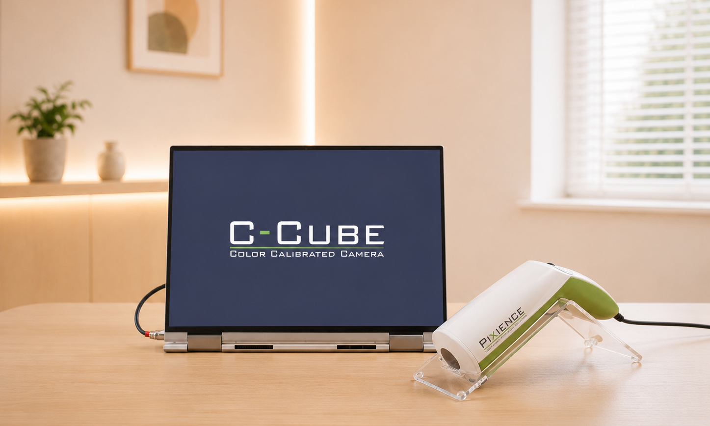
Thanks to its integrated mouse, you can navigate smoothly through the software while remaining close to your patient. Its USB 3 cable (3m) enables rapid installation and mobility to suit your practice.

Equipped with only three buttons, the C-Cube combines simplicity and efficiency, for intuitive handling. Designed to last, it has passed safety and resistance tests, ensuring reliability and longevity.

Software adapted to the rhythm of your consultations

The dermatology software associated with C-Cube allows you to simplify consultation management. Record and organize patient files, capture and analyze dermoscopic images with precision, track the evolution of lesions and generate detailed reports.

Intuitive and secure, it optimizes your workflow and follow-up quality.

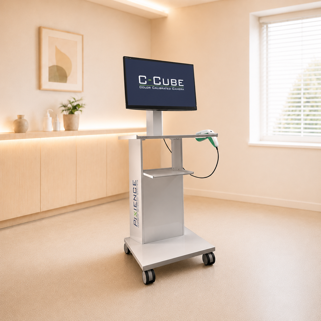
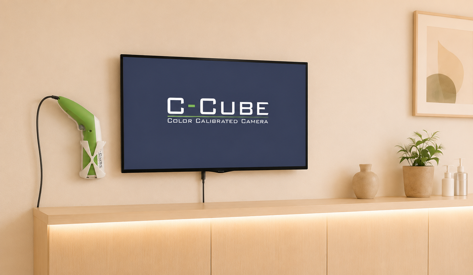
How to set up your practice with the C-Cube?

  • Resolution:  55 lpmm
  • Pixels: 18Mpx ou 12Mpx
  • Optical zoom: x20 to x80
  •  Digital zoom: x80 to x160
  • Smallest visible structure: 8µm
  • Image format: jpeg and DICOM
  • Power supply: USB 3.0 (5V, 2A)
  • Examination surface: 14.5 x 19.5 mm²
  • Weight: 410g